Blog

PCCN Exam Pancreatitis

PCCN Exam Pancreatitis

PCCN Exam Pancreatitis 

Pancreatitis

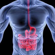
The pancreas is a large gland behind the stomach and next to the small intestine.  The pancreas does two things: it releases powerful digestive enzymes into the small intestine to aid the digestion of food, and it releases the hormones insulin and glucagon into the bloodstream.  These hormones help the body control how it uses food for energy. Pancreatitis is a disease in which the pancreas becomes inflamed.  Pancreatic damage happens when the digestive enzymes are activated before they are released into the small intestine and begin attacking the pancreas.  There are two forms of pancreatitis; acute and chronic.
  • Acute pancreatitis is a sudden inflammation that lasts for a short time.  It may range from mild discomfort to a severe life threatening illness.  Most people with acute pancreatitis recover completely after getting the right treatment.  In severe cases, acute pancreatitis can result in bleeding into the gland, serious tissue damage, infection, and cyst formation.
  • Chronic pancreatitis is long lasting inflammation of the pancreas.  it most often happens after an episode of acute pancreatitis.  Heavy alcohol consumption is another big cause.

Signs and Symptoms

  • Upper abdominal pain that radiates to the back; aggravated by eating, especially foods high in fat
  • Swollen and tender abdomen
  • Nausea and vomiting
  • Fever
  • Increased heart rate

Pancreatitis Etiology

  • Gallstones or heavy alcohol use
  • Medications, infections, trauma
  • Metabolic disorders and surgery

Treatment of Pancreatitis

  • Treat the s/s
  • IV fluids and pain meds

GI Sample Questions

1) Vasopressin may be used in the patients with GI bleeding. What is the mechanism of action of Vasopressin?

A) Increases mesenteric blood flow to reduce ischemia B) Decreases portal venous blood flow to decrease bleeding C) Causes sodium and water retention to replace volume D) Blocks H2 receptors to inhibit hydrochloric acid secretion

2) A 39 y/o male is admitted with a history of chronic liver failure and ETOH abuse. He has ascites and severe peripheral edema. He is anorexic, vomiting, hypokalemic, and now has developed metabolic alkalosis. Which of the following would not be included in this patient's management?

A) Diuretics B) Potassium supplements C) Antiemetics D) Diet high in protein

3) A 52 y/o male patient with acute pancreatitis develops agitation, fine tremors, and tachycardia about 48 hours after admission. Which of the following is the most likely cause of these signs and symptoms?

A) Pancreatic pseudocyst B) Hypoglycemia C) Alcohol withdrawal D) Pancreatic abscess


 GI Practice Questions Answer with Rationale

1) Correct Answer - B) Decreases portal venous blood flow to decrease bleeding
  • Rationale - Vasopressin slows blood loss by constricting the splanchnic arteriolar bed and decreasing portal venous pressure.
2) Correct Answer - A) Diuretics
  • Rationale - Diuretics would contribute to metabolic alkalosis and hypokalemia and would deplete the vascular bed rather than the third spaces.
3) Correct Answer - C) Alcohol withdrawal
  • Rationale - Alcoholism is a common cause of acute pancreatitis, and alcohol withdrawal is a complication that must be closely observed for within the first 24 - 72 hours after onset of abstinence.
 

PCCN National Exam Courses

Overview

  • Elite Reviews Offers A Variety Of Online Courses That Will More Than Adequately Help Prepare The Critical Care Nurse To Pass The National Exam.
  • Each Course Includes Continuing Education Credit and Sample Questions.

Continuing Education

  • Each Of Our Online Courses Has Been Approved Continuing Education Contact Hours by the California Board of Nursing
  • Login To Your Account In Order To Access The Course Completion Certificate Once The Course Is Complete.
PCCN Free Trial
  • FREE Sample Lecture & Practice Questions
  • Available For 24 Hrs After Registration
  • Click The Free Trial Link To Get Started - PCCN Free Trial


How It Works

How The Course Works

  • First - Purchase The Course By Clicking On The Blue Add To Cart Button - You Will Then Be Prompted To Create A User Account.
  • Second - After Creating An Account, All 3 Options (90, 120 or 150 Days) Will Be Listed. Select The Option You Desire And Delete The Other Two.
  • Third - You Will Be Prompted To Pay For The Review Using PayPal - After Payment You Will Be Redirected Back To Your Account.
  • Last - Click The Start Button Located Within Your Account To Begin The Program

PCCN Predictor Exam

PCCN Predictor Exam

  • 125 Prep Questions
  • Q & A With Rationales
  • Approved For 5 CEU's
  • 90 Days Availability
  • Cost $75.00


PCCN Question Bank

PCCN Question Bank

  • 1250+ Prep Questions
  • Q & A With Rationales
  • Approved For 25 CEU's
  • 90 Days Availability
  • Cost $200.00


PCCN Practice Questions

PCCN Practice Questions Bundle

  • 1350+ Prep Questions
  • Q & A With Rationales
  • Approved For 30 CEU's
  • 90 Days Availability
  • Cost $225.00


PCCN Review

PCCN Review Course 

  • Option 1
  • Lectures & 1250+ Questions
  • Q & A With Rationales
  • Approved For 35 CEU's
  • 90 Days Availability
  • Cost $275.00


PCCN Online Review

PCCN Online Review

  • Option 2
  • Lectures & 2000+ Questions
  • Q & A With Rationales
  • Approved For 40 CEU's
  • 90 Days Availability
  • Cost $325.00


PCCN Online Review

PCCN Review Course Bundle

  • Option 3
  • Lectures & 3000+ Questions
  • Q & A With Rationales
  • Approved For 70 CEU's
  • 90 Days Availability
  • Cost $375.00


Share On Social Media

Share On Social Media
 Connect With Us - Facebook - Instagram - Twitter 
Read more...

PCCN Exam AV Malformation

PCCN Exam AV Malformation

PCCN Exam AV Malformation

A brain arteriovenous malformation (AVM) is a tangle of abnormal blood vessels connecting arteries and veins in the brain.  The arteries are responsible for taking oxygen rich blood from the heart to the brain.  Veins carry the oxygen depleted blood back to the lungs and heart.  A brain AVM disrupts this vital process.  An AVM can develop anywhere in your body but occurs most often in the brain or spine.  Even so, brain AVM's are rare and affect less than 1% of the population.  Some of the causes of brain AVM is unknown, but researchers believe most brain AVMs emerge during fetal development. A brain AVM may not cause any signs or symptoms until the AVM ruptures, resulting in bleeding in the brain (hemorrhage).  In about half of all brain AVMs, hemorrhage is the first sign.  Some of the signs and symptoms are listed below...
  • Seizures
  • Headache
  • Muscle weakness or numbness in one part of the body
  • Vision loss
  • Difficulty speaking
  • Confusion
  • Severe unsteadiness

Risk Factors for AVM

Anyone can be born with a brain AVM, but these risk factors may include
  • Males - AVM's are more common in males
  • Family history

Complications 

  • Bleeding in the brain (hemorrhage)
  • Reduced oxygen to brain tissue
  • Thin or weak blood vessels
  • Brain damage

Diagnosis 

  • Cerebral arteriography
  • CT scan of the head
  • MRI of the brain

Treatment

  • Surgical removal (resection)
  • Endovascular embolization
  • Stereotactic Radiosurgery (SRS)

PCCN Exam - Practice Questions

1) A patient with a traumatic brain injury (TBI) has just been admitted after resuscitation in the emergency room department. Which of the following initial studies should the nurse anticipate in this patient?

A) Magnetic resonance imaging (MRI) of the brain B) Computed tomography (CT) scan of the head C) Lumbar puncture (LP) D) Cerebral angiography

2) A patient presents with new-onset seizures. Diagnostic imaging reveals a 4 cm right posterior frontal arteriovenous malformation. In planning this patient's nursing care needs, which of the following conditions should the nurse anticipate?

A) Left-sided weakness B) Receptive aphasia C) Left homonymous hemianopsia D) Sensory deficits on the right face and arm

3) A patient admitted last night with subarachnoid hemorrhage returned from the operating room 4 hours ago following aneurysm clipping. On admission, the patient was assessed as a Hunt and Hess Grade II. The patient now appears to be stuporous with significant left hemiparesis. The nurse should

A) Note that this is consistent with the Hunt and Hess appraisal at admission B) Contact the neurosurgeon to communicate the examination findings C) Adjust IV infusion rate because the aneurysm may have rebleed D) Prepare for ventriculostomy insertion by the physician so hydrocephalus can be treated.

4) During report, the nurse is told that a patient with an intracranial hemorrhage has GCS values of 3 - 3 - 4. Evaluation of the patient's progress is based on the nurse's knowledge that this patient

A) Opens his eyes when spoke to B) Follows simple commands C) Makes no attempt to remove noxious stimuli D) Makes no attempt to vocalize

5) A 25 y/o admitted patient has a generalized tonic clonic seizure. After the seizure has subsided, the nurse expects the patient to exhibit which of the following as a characteristic behavior after a seizure?

A) Restlessness B) Lethargy C) Automatisms D) Incontinence

PCCN Practice Questions Answer with Rationale

1) Correct Answer - B) Computed tomography (CT) scan of the head
  • Rationale - CT scan of the head is useful for looking at bone and blood and is the best imaging study to view most intracranial processes, including trauma, intracerebral hemorrhage, and hydrocephalus.
2) Correct Answer - A) Left-sided weakness
  • Rationale - The location of the AVM suggests which deficit the nurse needs to anticipate. A lesion in the right frontal area would be expected to affect voluntary motor control on the left side of the body. Comprehension of spoken language.
3) Correct Answer - B) Contact the neurosurgeon to communicate the examination findings
  • Rationale - The patient is now a Hunt and Hess Grade IV, a substantial deterioration compared to the initial Grade II (Grades range from I to V, with Grade I identifying alert patients with mild headache and stiff neck and Grade V identifying patients in coma. The most appropriate action would be to notify the neurosurgeon, who may request a head CT scan or angiography.
4) Correct Answer - A) Opens his eyes when spoke to
  • Rationale - The GCS is an internationally recognized standardized assessment tool that evaluates level of consciousness, the most sensitive indicator of cerebral function. The patient's best responses in three areas, eye opening, motor response, and verbal response.
5) Correct Answer - B) Lethargy
  • Rationale - Typical postictal behavior includes confusion, lethargy, HA, and somnolence. The patient will usually sleep for an extended period following a seizure.
 

PCCN National Exam Courses

Overview

  • Elite Reviews Offers A Variety Of Online Courses That Will More Than Adequately Help Prepare The Critical Care Nurse To Pass The National Exam.
  • Each Course Includes Continuing Education Credit and Sample Questions.

Continuing Education

  • Each Of Our Online Courses Has Been Approved Continuing Education Contact Hours by the California Board of Nursing
  • Login To Your Account In Order To Access The Course Completion Certificate Once The Course Is Complete.
PCCN Free Trial
  • FREE Sample Lecture & Practice Questions
  • Available For 24 Hrs After Registration
  • Click The Free Trial Link To Get Started - PCCN Free Trial


How It Works

How The Course Works

  • First - Purchase The Course By Clicking On The Blue Add To Cart Button - You Will Then Be Prompted To Create A User Account.
  • Second - After Creating An Account, All 3 Options (90, 120 or 150 Days) Will Be Listed. Select The Option You Desire And Delete The Other Two.
  • Third - You Will Be Prompted To Pay For The Review Using PayPal - After Payment You Will Be Redirected Back To Your Account.
  • Last - Click The Start Button Located Within Your Account To Begin The Program

PCCN Predictor Exam

PCCN Predictor Exam

  • 125 Prep Questions
  • Q & A With Rationales
  • Approved For 5 CEU's
  • 90 Days Availability
  • Cost $75.00


PCCN Question Bank

PCCN Question Bank

  • 1250+ Prep Questions
  • Q & A With Rationales
  • Approved For 25 CEU's
  • 90 Days Availability
  • Cost $200.00


PCCN Practice Questions

PCCN Practice Questions Bundle

  • 1350+ Prep Questions
  • Q & A With Rationales
  • Approved For 30 CEU's
  • 90 Days Availability
  • Cost $225.00


PCCN Review

PCCN Review Course 

  • Option 1
  • Lectures & 1250+ Questions
  • Q & A With Rationales
  • Approved For 35 CEU's
  • 90 Days Availability
  • Cost $275.00


PCCN Online Review

PCCN Online Review

  • Option 2
  • Lectures & 2000+ Questions
  • Q & A With Rationales
  • Approved For 40 CEU's
  • 90 Days Availability
  • Cost $325.00


PCCN Online Review

PCCN Review Course Bundle

  • Option 3
  • Lectures & 3000+ Questions
  • Q & A With Rationales
  • Approved For 70 CEU's
  • 90 Days Availability
  • Cost $375.00


Share On Social Media

Share On Social Media
 Connect With Us - Facebook - Instagram - Twitter 
Read more...

NCLEX Nephrotic Syndrome

NCLEX Nephrotic Syndrome Review

NCLEX Nephrotic Syndrome

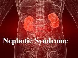
Nephrotic syndrome is a syndrome comprising signs of nephrosis, chiefly proteinuria, hypoalbuminemia, and edema.  It is a component of glomerulonephrosis, in which different degrees of proteinuria occur.  Essentially, loss of protein through the kidneys (proteinuria) leads to low protein levels in the blood (hypoproteinemia including hypoalbuminemia), which causes water to be drawn into soft tissues (edema). Very low hypoalbuminemia can also cause a variety of secondary problems, such as water in the abdominal cavity (ascites), around the heart or lung (pericardial effusion, pleural effusion), high cholesterol (hence hyperlipidemia), loss of molecules regulating coagulation (hence increased risk of thrombosis). Large proteinuria is due to an increase in permeability of the filtering membrane of the kidney which normally separates the blood from the urinary space in Bowman's capsule. This is composed of the capillary walls of the glomerulus which are wrapped by highly specialized cells called podocytes. Alterations in their capacity to filter the substances transported in the blood mean that proteins but not cells pass into the urine (hence no hematuria). By contrast, in nephritic syndrome red blood cells pass through the pores, causing hematuria.

Signs and Symptoms

Nephrotic syndrome is characterized by large proteinuria, hypoalbuminemia, hyperlipidemia, and edema (which is generalized and also known as anasarca or dropsy) that begins in the face. Lipiduria (lipids in urine) can also occur, but is not essential for the diagnosis of nephrotic syndrome. Hyponatremia also occurs with a low fractional sodium excretion. Hyperlipidemia is caused by two factors:
  • Hypoproteinemia stimulates protein synthesis in the liver, resulting in the overproduction of lipoproteins.
  • Lipid catabolism is decreased due to lower levels of lipoprotein lipase, the main enzyme involved in lipoprotein breakdown.  Cofactors, such as apolipoprotein C2 may also be lost by increased filtration of proteins.
A few other characteristics seen in nephrotic syndrome are:
  • The most common sign is excess fluid in the body due to the serum hypoalbuminemia. Lower serum oncotic pressure causes fluid to accumulate in the interstitial tissues. Sodium and water retention aggravates the edema. This may take several forms:
    • Puffiness around the eyes, characteristically in the morning.
    • Pitting edema over the legs.
    • Fluid in the pleural cavity causing pleural effusion. More commonly associated with excess fluid is pulmonary edema.
    • Fluid in the peritoneal cavity causing ascites.
    • Generalized edema throughout the body known as anasarca.
  • Most of the patients are normotensive but hypertension (rarely) may also occur.
  • Anemia (iron resistant microcytic hypochromic type) maybe present due to transferrin loss.
  • Dyspnea may be present due to pleural effusion or due to diaphragmatic compression with ascites.
  • Erythrocyte sedimentation rate is increased due to increased fibrinogen & other plasma contents.
  • Some patients may notice foamy or frothy urine, due to a lowering of the surface tension by the severe proteinuria. Actual urinary complaints such as hematuria or oliguria are uncommon, though these are seen commonly in nephritic syndrome.
  • May have features of the underlying cause, such as the rash associated with systemic lupus erythematosus, or the neuropathy associated with diabetes.

Causes

Nephrotic syndrome is usually caused by damage to the clusters of tiny blood vessels (glomeruli) of your kidneys. The glomeruli filter your blood as it passes through your kidneys, separating things your body needs from those it doesn't. Healthy glomeruli keep blood protein (mainly albumin) — which is needed to maintain the right amount of fluid in your body — from seeping into your urine. When damaged, glomeruli allow too much blood protein to leave your body, leading to nephrotic syndrome. Many diseases and conditions can cause glomerular damage and lead to nephrotic syndrome, including:
  • Minimal change disease. The most common cause of nephrotic syndrome in children, this disorder results in abnormal kidney function, but when the kidney tissue is examined under a microscope, it appears normal or nearly normal. The cause of the abnormal function typically can't be determined.
  • Focal segmental glomerulosclerosis. Characterized by scattered scarring of some of the glomeruli, this condition may result from another disease or a genetic defect or occur for no known reason.
  • Membranous nephropathy. This kidney disorder is the result of thickening membranes within the glomeruli. The exact cause of the thickening isn't known, but it's sometimes associated with other medical conditions, such as hepatitis B, malaria, lupus and cancer.
  • Diabetic kidney disease. Diabetes can lead to kidney damage (diabetic nephropathy) that affects the glomeruli.
  • Systemic lupus erythematosus. This chronic inflammatory disease can lead to serious kidney damage.
  • Amyloidosis. This disorder occurs when substances called amyloid proteins accumulate in your organs. Amyloid buildup often affects the kidneys, damaging their filtering system.
  • Blood clot in a kidney vein. Renal vein thrombosis, which occurs when a blood clot blocks a vein connected to the kidney, can cause nephrotic syndrome.
  • Heart failure. Some forms of heart failure, such as constrictive pericarditis and severe right heart failure, can cause nephrotic syndrome.

Risk Factors

Factors that can increase your risk of nephrotic syndrome include:
  • Medical conditions that can damage your kidneys. Certain diseases and conditions increase your risk of developing nephrotic syndrome, such as diabetes, lupus, amyloidosis, minimal change disease and other kidney diseases.
  • Certain medications. Examples of medications that can cause nephrotic syndrome include nonsteroidal anti-inflammatory drugs and drugs used to fight infections.
  • Certain infections. Examples of infections that increase the risk of nephrotic syndrome include HIV, hepatitis B, hepatitis C and malaria.

Complications

Possible complications of nephrotic syndrome include:
  • Blood clots. The inability of the glomeruli to filter blood properly can lead to loss of blood proteins that help prevent clotting. This increases your risk of developing a blood clot (thrombus) in your veins.
  • High blood cholesterol and elevated blood triglycerides. When the level of the protein albumin in your blood falls, your liver makes more albumin. At the same time, your liver releases more cholesterol and triglycerides.
  • Poor nutrition. Loss of too much blood protein can result in malnutrition. This can lead to weight loss, but it may be masked by swelling. You may also have too few red blood cells (anemia) and low levels of vitamin D and calcium.
  • High blood pressure. Damage to your glomeruli and the resulting buildup of wastes in your bloodstream (uremia) can raise your blood pressure.
  • Acute kidney failure. If your kidneys lose their ability to filter blood due to damage to the glomeruli, waste products may build up quickly in your blood. If this happens, you may need emergency dialysis — an artificial means of removing extra fluids and waste from your blood — typically with an artificial kidney machine (dialyzer).
  • Chronic kidney disease. Nephrotic syndrome may cause your kidneys to gradually lose their function over time. If kidney function falls low enough, you may require dialysis or a kidney transplant.
  • Infections. People with nephrotic syndrome have an increased risk of infections.

Treatments

Treatment for nephrotic syndrome involves treating any underlying medical condition that may be causing your nephrotic syndrome. Your doctor may also recommend medications that may help control your signs and symptoms or treat complications of nephrotic syndrome. Medications may include:
    • Blood pressure medications
    • Diuretics help control swelling by increasing your kidneys' fluid output. Diuretic medications include furosemide (Lasix) and spironolactone (Aldactone).
    • Cholesterol-reducing medications. Medications called statins can help lower cholesterol levels. However, it's currently unclear whether or not cholesterol-lowering medications can specifically improve the outcomes of people with nephrotic syndrome, such as avoiding heart attacks or decreasing the risk of early death. Statins include atorvastatin (Lipitor), fluvastatin (Lescol), lovastatin (Altoprev, Mevacor), pravastatin (Pravachol), rosuvastatin (Crestor) and simvastatin (Zocor).
    • Blood thinners. Medications called anticoagulants help decrease your blood's ability to clot and reduce your risk of developing blood clots. Anticoagulants include heparin or warfarin (Coumadin).
    • Immune system-suppressing medications. Medications to control the immune system, such as corticosteroids, may decrease the inflammation that accompanies certain kidney disorders, such as minimal change disease.
 

NCLEX National Exam Courses

Overview

  • Elite Reviews Offers A Variety Of Online Courses That Will More Than Adequately Help Prepare The Graduate Nurse To Pass The National Exam.
  • Each Course Includes Sample Questions & The Most Current NCLEX Exam Updates.
NCLEX Free Trial
  • FREE Sample Lecture & Practice Questions
  • Available For 24 Hrs After Registration
  • Click The Free Trial Link To Get Started - NCLEX Free Trial


How It Works

How The Course Works

  • First - Purchase The Course By Clicking On The Blue Add To Cart Button - You Will Then Be Prompted To Create A User Account.
  • Second - After Creating An Account, All 3 Options (90, 120, 150 Days) Will Be Listed. Select The Option You Desire And Delete The Other Two.
  • Third - You Will Be Prompted To Pay For This Review Using PayPal - After Payment You Will Be Redirected Back To Your Account.
  • Last - Click The Start Button Located Within Your Account To Begin The Course

NCLEX Predictor Exam

NCLEX Predictor Exam

  • 175 Prep Questions
  • Q & A With Rationales
  • Alt. Format Questions
  • 90 Days Availability
  • Cost $75.00


NCLEX Question Bank

NCLEX Question Bank

  • 1250+ Prep Questions
  • Q & A With Rationales
  • Alt. Format Questions
  • 90 Days Availability
  • Cost $200.00


NCLEX Practice Questions

NCLEX Practice Questions Bundle

  • 1350+ Prep Questions
  • Q & A With Rationales
  • Alt. Format Questions
  • 90 Days Availability
  • Cost $225.00


NCLEX Review

NCLEX Review Course

  • Option 1
  • Lectures & 1250+ Questions
  • Q & A With Rationales
  • Alt. Format Questions
  • 90 Days Availability
  • Cost $275.00


NCLEX Online Review

NCLEX Online Review

  • Option 2
  • Lectures & 2000+ Questions
  • Q & A With Rationales
  • Alt. Format Questions
  • 90 Days Availability
  • Cost $325.00


NCLEX Online Review

NCLEX Review Course Bundle

  • Option 3
  • Lectures & 3000+ Questions
  • Q & A With Rationales
  • Alt. Format Questions
  • 90 Days Availability
  • Cost $375.00


Share On Social Media

Share On Social Media
 Connect With Us - Facebook - Instagram - Twitter 
Read more...

NCLEX Glomerulonephritis

NCLEX Glomerulonephritis Review

NCLEX Glomerulonephritis

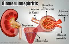
Glomerulonephritis is inflammation of the tiny filters in your kidneys (glomeruli). Glomeruli remove excess fluid, electrolytes and waste from your bloodstream and pass them into your urine. Also called glomerular disease, glomerulonephritis can be acute — a sudden attack of inflammation — or chronic — coming on gradually. Glomerulonephritis (GN), also known as glomerular nephritis, is a term used to refer to several kidney diseases (usually affecting both kidneys). Many of the diseases are characterized by inflammation either of the glomeruli or of the small blood vessels in the kidneys, hence the name, but not all diseases necessarily have an inflammatory component. As it is not strictly a single disease, its presentation depends on the specific disease entity: it may present with isolated hematuria and/or proteinuria (blood or protein in the urine); or as a nephrotic syndrome, a nephritic syndrome, acute kidney injury, or chronic kidney disease. They are categorized into several different pathological patterns, which are broadly grouped into non-proliferative or proliferative types. Diagnosing the pattern of GN is important because the outcome and treatment differs in different types. Primary causes are intrinsic to the kidney. Secondary causes are associated with certain infections (bacterial, viral or parasitic pathogens), drugs, systemic disorders (SLE, vasculitis), or diabetes.

Signs and Symptoms

Signs and symptoms of glomerulonephritis depend on whether you have the acute or chronic form, and the cause. Your first indication that something is wrong may come from symptoms or from the results of a routine urinalysis. Glomerulonephritis signs and symptoms may include:
  • Pink or cola-colored urine from red blood cells in your urine (hematuria)
  • Foamy urine due to excess protein (proteinuria)
  • High blood pressure (hypertension)
  • Fluid retention (edema) with swelling evident in your face, hands, feet and abdomen
  • Fatigue from anemia or kidney failure

Causes

Infections

  • Post-streptococcal glomerulonephritis. Glomerulonephritis may develop a week or two after recovery from a strep throat infection or, rarely, a skin infection (impetigo). To fight the infection, your body produces extra antibodies that may eventually settle in the glomeruli, causing inflammation. Children are more likely to develop post-streptococcal glomerulonephritis than are adults, and they're also more likely to recover quickly.
  • Bacterial endocarditis. Bacteria occasionally can spread through your bloodstream and lodge in your heart, causing an infection of one or more of your heart valves. You're at greater risk of this condition if you have a heart defect, such as a damaged or artificial heart valve. Bacterial endocarditis is associated with glomerular disease, but the exact connection between the two is unclear.
  • Viral infections. Viral infections, such as the human immunodeficiency virus (HIV), hepatitis B and hepatitis C, may trigger glomerulonephritis.

 Immune diseases

  • Lupus. A chronic inflammatory disease, lupus can affect many parts of your body, including your skin, joints, kidneys, blood cells, heart and lungs.
  • Goodpasture's syndrome. A rare immunological lung disorder that may mimic pneumonia, Goodpasture's syndrome causes bleeding in your lungs as well as glomerulonephritis.
  • IgA nephropathy. Characterized by recurrent episodes of blood in the urine, this primary glomerular disease results from deposits of immunoglobulin A (IgA) in the glomeruli. IgA nephropathy can progress for years with no noticeable symptoms.

Vasculitis

  • Polyarteritis. This form of vasculitis affects small and medium blood vessels in many parts of your body, such as your heart, kidneys and intestines.
  • Wegener's granulomatosis. This form of vasculitis affects small and medium blood vessels in your lungs, upper airways and kidneys.

Conditions likely to cause scarring of the glomeruli

  • High blood pressure. High blood pressure can damage your kidneys and impair their ability to function normally. Glomerulonephritis can also lead to high blood pressure because it reduces kidney function and may influence how your kidneys handle sodium.
  • Diabetic kidney disease. Diabetic kidney disease (diabetic nephropathy) can affect anyone with diabetes. Diabetic nephropathy usually takes years to develop. Good control of blood sugar levels and blood pressure may prevent or slow kidney damage.
  • Focal segmental glomerulosclerosis. Characterized by scattered scarring of some of the glomeruli, this condition may result from another disease or occur for no known reason.
Chronic glomerulonephritis sometimes develops after a bout of acute glomerulonephritis. In some people there's no history of kidney disease, so the first indication of chronic glomerulonephritis is kidney failure. Infrequently, chronic glomerulonephritis runs in families. One inherited form, Alport syndrome, also may involve hearing or vision impairment.

Complications

  • Acute kidney failure. Loss of function in the filtering part of the nephron may cause waste products to accumulate rapidly. You may need emergency dialysis — an artificial means of removing extra fluids and waste from your blood — typically by an artificial kidney machine.
  • Chronic kidney disease. With chronic kidney disease, your kidneys gradually lose their filtering ability. If your kidney function deteriorates to less than 10 percent of normal capacity, you have end-stage kidney disease, which requires dialysis or a kidney transplant to sustain life.
  • High blood pressure. Damage to your kidneys and the resulting buildup of wastes in the bloodstream can raise your blood pressure.
  • Nephrotic syndrome. With nephrotic syndrome, you have too much protein in your urine, which results in too little protein in your blood. Nephrotic syndrome may also be associated with high blood cholesterol and swelling (edema) of the eyelids, feet and abdomen.

Treatments

Treatment of glomerulonephritis and your outcome depend on:
  • Whether you have an acute or chronic form of the disease
  • The underlying cause
  • The type and severity of your signs and symptoms
Treat high blood pressure and underlying cause  

NCLEX National Exam Courses

Overview

  • Elite Reviews Offers A Variety Of Online Courses That Will More Than Adequately Help Prepare The Graduate Nurse To Pass The National Exam.
  • Each Course Includes Sample Questions & The Most Current NCLEX Exam Updates.
NCLEX Free Trial
  • FREE Sample Lecture & Practice Questions
  • Available For 24 Hrs After Registration
  • Click The Free Trial Link To Get Started - NCLEX Free Trial


How It Works

How The Course Works

  • First - Purchase The Course By Clicking On The Blue Add To Cart Button - You Will Then Be Prompted To Create A User Account.
  • Second - After Creating An Account, All 3 Options (90, 120, 150 Days) Will Be Listed. Select The Option You Desire And Delete The Other Two.
  • Third - You Will Be Prompted To Pay For This Review Using PayPal - After Payment You Will Be Redirected Back To Your Account.
  • Last - Click The Start Button Located Within Your Account To Begin The Course

NCLEX Predictor Exam

NCLEX Predictor Exam

  • 175 Prep Questions
  • Q & A With Rationales
  • Alt. Format Questions
  • 90 Days Availability
  • Cost $75.00


NCLEX Question Bank

NCLEX Question Bank

  • 1250+ Prep Questions
  • Q & A With Rationales
  • Alt. Format Questions
  • 90 Days Availability
  • Cost $200.00


NCLEX Practice Questions

NCLEX Practice Questions Bundle

  • 1350+ Prep Questions
  • Q & A With Rationales
  • Alt. Format Questions
  • 90 Days Availability
  • Cost $225.00


NCLEX Review

NCLEX Review Course

  • Option 1
  • Lectures & 1250+ Questions
  • Q & A With Rationales
  • Alt. Format Questions
  • 90 Days Availability
  • Cost $275.00


NCLEX Online Review

NCLEX Online Review

  • Option 2
  • Lectures & 2000+ Questions
  • Q & A With Rationales
  • Alt. Format Questions
  • 90 Days Availability
  • Cost $325.00


NCLEX Online Review

NCLEX Review Course Bundle

  • Option 3
  • Lectures & 3000+ Questions
  • Q & A With Rationales
  • Alt. Format Questions
  • 90 Days Availability
  • Cost $375.00


Share On Social Media

Share On Social Media
 Connect With Us - Facebook - Instagram - Twitter 
Read more...

NCLEX Coarctation of the Aorta

NCLEX Coarctation of the Aorta

NCLEX Coarctation Of The Aorta

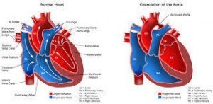
Coarctation of the aorta is a birth defect in which a part of the aorta is narrower than usual. If the narrowing is severe enough and if it is not diagnosed, the baby may have serious problems and may need surgery or other procedures soon after birth. For this reason, coarctation of the aorta is often considered a critical congenital heart defect. The defect occurs when a baby’s aorta does not form correctly as the baby grows and develops during pregnancy. The narrowing of the aorta usually happens in the part of the blood vessel just after the arteries branch off to take blood to the head and arms, near the patent ductus arteriosus, although sometimes the narrowing occurs before or after the ductus arteriosus. In some babies with coarctation, it is thought that some tissue from the wall of ductus arteriosus blends into the tissue of the aorta. When the tissue tightens and allows the ductus arteriosus to close normally after birth, this extra tissue may also tighten and narrow the aorta. The narrowing, or coarctation, blocks normal blood flow to the body. This can back up flow into the left ventricle of the heart, making the muscles in this ventricle work harder to get blood out of the heart. Since the narrowing of the aorta is usually located after arteries branch to the upper body, coarctation in this region can lead to normal or high blood pressure and pulsing of blood in the head and arms and low blood pressure and weak pulses in the legs and lower body. If the condition is very severe, enough blood may not be able to get through to the lower body. The extra work on the heart can cause the walls of the heart to become thicker in order to pump harder. This eventually weakens the heart muscle. If the aorta is not widened, the heart may weaken enough that it leads to heart failure. Coarctation of the aorta often occurs with other congenital heart defects.

Signs and Symptoms

Coarctation of the aorta symptoms depend on the severity of the condition. Most people don't have symptoms. Children with serious aortic narrowing may show signs and symptoms earlier in life, but mild cases with no symptoms might not be diagnosed until adulthood. People may also have signs or symptoms of other heart defects that they have along with coarctation of the aorta.
Babies with severe coarctation of the aorta may begin having signs and symptoms shortly after birth. These include:
  • Pale skin
  • Irritability
  • Heavy sweating
  • Difficulty breathing
  • Difficulty feeding

Causes

The causes of heart defects, including coarctation of the aorta, among most babies are unknown. Some babies have heart defects because of changes in their genes or chromosomes. Heart defects, like coarctation of the aorta, are also thought to be caused by a combination of genes and other risk factors, such as things the mother comes in contact with in the environment, what the mother eats or drinks, or medicines the mother uses.

Complications

Untreated coarctation of the aorta frequently leads to complications. Several of the complications may be a result of long-standing high blood pressure caused by the aortic coarctation. Complications are also possible after treatment for coarctation of the aorta. Complications of coarctation of the aorta may include:
  • High blood pressure
  • Stroke
  • Enlargement in a section of the wall of the aorta (aneurysm)
  • Aortic rupture or tear (dissection)
  • Premature coronary artery disease — narrowing of the blood vessels that supply the heart
  • Heart failure
  • A weakened or bulging artery in the brain (brain aneurysm) or bleeding in the brain (hemorrhage)

Treatments

Treatment options for coarctation of the aorta depend on your age at the time of diagnosis and the severity of your condition. Other heart defects might be repaired at the same time as aortic coarctation. Treatment approaches usually consist of surgery or a procedure called balloon angioplasty.  

NCLEX National Exam Courses

Overview

  • Elite Reviews Offers A Variety Of Online Courses That Will More Than Adequately Help Prepare The Graduate Nurse To Pass The National Exam.
  • Each Course Includes Sample Questions & The Most Current NCLEX Exam Updates.
NCLEX Free Trial
  • FREE Sample Lecture & Practice Questions
  • Available For 24 Hrs After Registration
  • Click The Free Trial Link To Get Started - NCLEX Free Trial


How It Works

How The Course Works

  • First - Purchase The Course By Clicking On The Blue Add To Cart Button - You Will Then Be Prompted To Create A User Account.
  • Second - After Creating An Account, All 3 Options (90, 120, 150 Days) Will Be Listed. Select The Option You Desire And Delete The Other Two.
  • Third - You Will Be Prompted To Pay For This Review Using PayPal - After Payment You Will Be Redirected Back To Your Account.
  • Last - Click The Start Button Located Within Your Account To Begin The Course

NCLEX Predictor Exam

NCLEX Predictor Exam

  • 175 Prep Questions
  • Q & A With Rationales
  • Alt. Format Questions
  • 90 Days Availability
  • Cost $75.00


NCLEX Question Bank

NCLEX Question Bank

  • 1250+ Prep Questions
  • Q & A With Rationales
  • Alt. Format Questions
  • 90 Days Availability
  • Cost $200.00


NCLEX Practice Questions

NCLEX Practice Questions Bundle

  • 1350+ Prep Questions
  • Q & A With Rationales
  • Alt. Format Questions
  • 90 Days Availability
  • Cost $225.00


NCLEX Review

NCLEX Review Course

  • Option 1
  • Lectures & 1250+ Questions
  • Q & A With Rationales
  • Alt. Format Questions
  • 90 Days Availability
  • Cost $275.00


NCLEX Online Review

NCLEX Online Review

  • Option 2
  • Lectures & 2000+ Questions
  • Q & A With Rationales
  • Alt. Format Questions
  • 90 Days Availability
  • Cost $325.00


NCLEX Online Review

NCLEX Review Course Bundle

  • Option 3
  • Lectures & 3000+ Questions
  • Q & A With Rationales
  • Alt. Format Questions
  • 90 Days Availability
  • Cost $375.00


Share On Social Media

Share On Social Media
 Connect With Us - Facebook - Instagram - Twitter 
Read more...

NCLEX Mitral Valve Prolapse

NCLEX Mitral Valve Prolapse Review

NCLEX Mitral Valve Prolapse

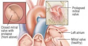
Mitral valve prolapse (MVP) (a.k.a. floppy mitral valve syndrome, systolic click murmur syndrome or billowing mitral leaflet) is a valvular heart disease characterized by the displacement of an abnormally thickened mitral valve leaflet into the left atrium during systole. It is the primary form of myxomatous degeneration of the valve. There are various types of MVP, broadly classified as classic and nonclassic. In its nonclassic form, MVP carries a low risk of complications and often can be kept minimal by dietary attention. In severe cases of classic MVP, complications include mitral regurgitation, infective endocarditis, congestive heart failure, and, in rare circumstances, cardiac arrest. The mitral valve, so named because of its resemblance to a bishop's mitre, is the heart valve that prevents the backflow of blood from the left ventricle into the left atrium of the heart. It is composed of two leaflets, one anterior and one posterior, that close when the left ventricle contracts. Each leaflet is composed of three layers of tissue: the atrialis, fibrosa, and spongiosa. Patients with classic mitral valve prolapse have excess connective tissue that thickens the spongiosa and separates collagen bundles in the fibrosa. This is due to an excess of dermatan sulfate, a glycosaminoglycan. This weakens the leaflets and adjacent tissue, resulting in increased leaflet area and elongation of the chordae tendineae. Elongation of the chordae tendineae often causes rupture, commonly to the chordae attached to the posterior leaflet. Advanced lesions—also commonly involving the posterior leaflet—lead to leaflet folding, inversion, and displacement toward the left atrium.

Signs and Symptoms

  • A racing or irregular heartbeat (arrhythmia)
  • Dizziness or lightheadedness
  • Difficulty breathing or shortness of breath, often when lying flat or during physical activity
  • Fatigue
  • Chest pain that's not caused by a heart attack or coronary artery disease

Causes

When your heart is working properly, the mitral valve closes completely during contraction of the left ventricle and prevents blood from flowing back into your heart's upper left chamber (left atrium). But in some people with mitral valve prolapse, one or both of the mitral valve's flaps (leaflets) have extra tissue bulging (prolapsing) like a parachute into the left atrium each time the heart contracts.
The bulging may keep the valve from closing tightly. When blood leaks backward through the valve, it's called mitral valve regurgitation.
This may not cause problems if only a small amount of blood leaks back into the atrium. More severe mitral valve regurgitation can cause symptoms such as shortness of breath, fatigue or lightheadedness. Another name for mitral valve prolapse is click-murmur syndrome. When a doctor listens to your heart using a stethoscope, he or she may hear a clicking sound as the valve's leaflets billow out, followed by a murmur resulting from blood flowing back into the atrium. Other names to describe mitral valve prolapse include:
  • Barlow's syndrome
  • Floppy valve syndrome
  • Balloon mitral valve
  • Billowing mitral valve
  • Myxomatous mitral valve
  • Prolapsing mitral valve syndrome

Risk Factors

Mitral valve prolapse can develop in any person at any age.  Serious symptoms of mitral valve prolapse tend to occur most often in men older than 50. Mitral valve prolapse can run in families and may be linked to several other conditions, such as:
  • Marfan syndrome
  • Ehlers-Danlos syndrome
  • Ebstein's anomaly
  • Muscular dystrophy
  • Graves' disease
  • Scoliosis

Complications 

Although most people with mitral valve prolapse never have problems, complications can occur. They may include:
  • Mitral valve regurgitation. The most common complication is a condition in which the valve leaks blood back into the left atrium (mitral valve regurgitation).
    Being male or having high blood pressure increases your risk of mitral valve regurgitation. If the regurgitation is severe, you may need surgery to repair or replace the valve in order to prevent the development of complications, such as stroke.
  • Heart rhythm problems (arrhythmias). Irregular heart rhythms can occur in people with mitral valve prolapse. These most commonly occur in the upper chambers of the heart, and while they may be bothersome, they aren't usually life-threatening. People with severe mitral valve regurgitation or severe deformity of their mitral valve are most at risk of having serious rhythm problems, which affect blood flow through the heart.
  • Heart valve infection (endocarditis). The inside of your heart contains four chambers and four valves lined by a thin membrane called the endocardium. Endocarditis is an infection of this inner lining. An abnormal mitral valve increases your chance of getting endocarditis from bacteria, which can further damage the mitral valve. The risk is higher in older men. Doctors used to recommend that some people with mitral valve prolapse take antibiotics before certain dental or medical procedures to prevent endocarditis but not anymore. The American Heart Association advises that antibiotics aren't necessary in most cases for someone with mitral valve regurgitation or mitral valve prolapse.

Treatments

Most people with mitral valve prolapse, particularly people without symptoms, don't require treatment. If you have mitral valve regurgitation but don't have symptoms, your doctor may suggest you return regularly for follow-up examinations to monitor your condition, depending on the severity of your condition. If you develop symptoms, your doctor might prescribe certain medications to treat mitral valve prolapse-related chest pain, heart rhythm abnormalities or other complications. Some medications you might be prescribed include:
  • Beta blockers
  • Diuretics
  • Heart rhythm medications
  • Aspirin
Surgery
  • Valve Replacement or Repair
 

NCLEX National Exam Courses

Overview

  • Elite Reviews Offers A Variety Of Online Courses That Will More Than Adequately Help Prepare The Graduate Nurse To Pass The National Exam.
  • Each Course Includes Sample Questions & The Most Current NCLEX Exam Updates.
NCLEX Free Trial
  • FREE Sample Lecture & Practice Questions
  • Available For 24 Hrs After Registration
  • Click The Free Trial Link To Get Started - NCLEX Free Trial


How It Works

How The Course Works

  • First - Purchase The Course By Clicking On The Blue Add To Cart Button - You Will Then Be Prompted To Create A User Account.
  • Second - After Creating An Account, All 3 Options (90, 120, 150 Days) Will Be Listed. Select The Option You Desire And Delete The Other Two.
  • Third - You Will Be Prompted To Pay For This Review Using PayPal - After Payment You Will Be Redirected Back To Your Account.
  • Last - Click The Start Button Located Within Your Account To Begin The Course

NCLEX Predictor Exam

NCLEX Predictor Exam

  • 175 Prep Questions
  • Q & A With Rationales
  • Alt. Format Questions
  • 90 Days Availability
  • Cost $75.00


NCLEX Question Bank

NCLEX Question Bank

  • 1250+ Prep Questions
  • Q & A With Rationales
  • Alt. Format Questions
  • 90 Days Availability
  • Cost $200.00


NCLEX Practice Questions

NCLEX Practice Questions Bundle

  • 1350+ Prep Questions
  • Q & A With Rationales
  • Alt. Format Questions
  • 90 Days Availability
  • Cost $225.00


NCLEX Review

NCLEX Review Course

  • Option 1
  • Lectures & 1250+ Questions
  • Q & A With Rationales
  • Alt. Format Questions
  • 90 Days Availability
  • Cost $275.00


NCLEX Online Review

NCLEX Online Review

  • Option 2
  • Lectures & 2000+ Questions
  • Q & A With Rationales
  • Alt. Format Questions
  • 90 Days Availability
  • Cost $325.00


NCLEX Online Review

NCLEX Review Course Bundle

  • Option 3
  • Lectures & 3000+ Questions
  • Q & A With Rationales
  • Alt. Format Questions
  • 90 Days Availability
  • Cost $375.00


Share On Social Media

Share On Social Media
 Connect With Us - Facebook - Instagram - Twitter 
Read more...

NCLEX Aortic Stenosis

 NCLEX Aortic Stenosis Review

NCLEX Aortic Stenosis

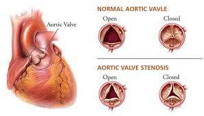
Aortic stenosis (AS or AOS) is the narrowing of the exit of the left ventricle of the heart, such that problems result. It may occur at the aortic valve as well as above and below this level. It typically gets worse over time. Symptoms often come on gradually with a decreased ability to exercise often occurring first. If heart failure, loss of consciousness, or heart related chest pain occurs due to AS the outcomes are worse. Loss of consciousness typically occurs with standing or exercise. Signs of heart failure include shortness of breath especially when lying down, at night, or with exercise, and swelling of the legs. Thickening of the valve without narrowing is known as aortic sclerosis.

Signs and Symptoms

Aortic valve stenosis ranges from mild to severe. Aortic valve stenosis signs and symptoms generally develop when narrowing of the valve is severe and can include:
  • Chest pain (angina) or tightness
  • Feeling faint or fainting with exertion
  • Shortness of breath, especially with exertion
  • Fatigue, especially during times of increased activity
  • Heart palpitations — sensations of a rapid, fluttering heartbeat
  • Heart murmur

Causes

  • As the aortic valve narrows, the left ventricle has to work harder to pump blood out through the valve. To do this extra work, the muscles in the ventricle walls become thicker. This can lead to chest pain.
  • As the pressure continues to rise, blood may back up into the lungs. Severe aortic stenosis can limit the amount of blood that reaches the brain and the rest of the body.
  • Aortic stenosis may be present from birth (congenital), but most often it develops later in life. Children with aortic stenosis may have other conditions present from birth.
  • Aortic stenosis mainly occurs due to the buildup of calcium deposits that narrow the valve. This is called calcific aortic stenosis. The problem mostly affects older people.
  • Calcification of the valve happens sooner in people who are born with abnormal aortic or bicuspid valves. In rare cases, calcification can develop more quickly when a person has received chest radiation (such as for cancer treatment).
  • Another cause is rheumatic fever. This condition can develop after strep throat or scarlet fever. Valve problems do not develop for 5 - 10 years or longer after rheumatic fever occurs. Rheumatic fever is becoming rarer in the United States.
  • Aortic stenosis occurs in about 2% of people over 65 years of age. It occurs more often in men than in women.

Risk Factors

Aortic valve stenosis isn't considered preventable, and presently it's not known why some people develop this condition. Some risk factors include:
  • A deformed aortic valve Some people are born with an already narrowed aortic valve or develop aortic valve stenosis later in life because they were born with a bicuspid aortic valve — one with two flaps (leaflets) instead of three. People may also develop aortic valve stenosis if they were born with one leaflet (unicuspid aortic valve) or four leaflets (quadricuspid aortic valve), but these are much more rare conditions.
    A bicuspid aortic valve is a major risk factor for aortic valve stenosis. A bicuspid aortic valve can run in families, so knowing your family history is important. If you have a first-degree relative — a parent, sibling or child — with a bicuspid aortic valve, it is reasonable to check to see if you have this abnormality.
  • Age Aortic valve stenosis may be related to increasing age and the buildup of calcium deposits on heart valves.
  • Previous rheumatic fever  Rheumatic fever can cause the flaps (leaflets) of your aortic valve to stiffen and fuse, eventually resulting in aortic valve stenosis.
  • Chronic kidney disease  Aortic valve stenosis is associated with chronic kidney disease.
Risk factors for aortic valve stenosis and atherosclerotic heart disease are similar — such as high blood pressure, high cholesterol, type 2 diabetes and smoking — which may indicate a link between the two.

Complications

Aortic valve stenosis — of any cause — can be a serious condition. If the aortic valve is narrowed, the left ventricle has to work harder to pump a sufficient amount of blood into the aorta and onward to the rest of your body. In response, the left ventricle may thicken and enlarge. At first, these adaptations help the left ventricle pump blood with more force. But eventually it's harder for the heart to maintain the blood flow to the body through the narrowed valve. Then you will start to experience symptoms. Eventually, the extra work of the heart can weaken the left ventricle — and your heart overall.
Left unchecked, aortic valve stenosis can lead to life-threatening heart problems, including:
  • Chest pain (angina)
  • Fainting (syncope)
  • Heart failure
  • Irregular heart rhythms (arrhythmias)
  • Cardiac arrest

Diagnosis

A heart murmur, click, or other abnormal sound is almost always heard through a stethoscope. The health care provider may be able to feel a vibration or movement when placing a hand over the heart. There may be a faint pulse or changes in the quality of the pulse in the neck. Blood pressure may be low. The following tests may also be performed:
  • ECG
  • ECHO
  • Chest X-Ray
  • CT Chest
  • Exercise stress testing
  • Left cardiac catheterization
  • MRI of the heart
  • Transesophageal echocardiogram (TEE)

Treatments

Treatment is generally not necessary in people without symptoms.  In moderate cases, echocardiography is performed every 1–2 years to monitor the progression, possibly complemented with a cardiac stress test.  In severe cases, echocardiography is performed every 3–6 months.  In both moderate and mild cases, the person should immediately make a revisit or be admitted for inpatient care if any new related symptoms appear.  There are no therapeutic options currently available to treat people with aortic valve stenosis; however, studies have indicated that the disease occurs as a result of active cellular processes, suggesting that targeting these processes may lead to viable therapeutic approaches.

Surgical Repair

  • Aortic Valve Repair and/or Replacement
  • Balloon valvuloplasty
 

NCLEX National Exam Courses

Overview

  • Elite Reviews Offers A Variety Of Online Courses That Will More Than Adequately Help Prepare The Graduate Nurse To Pass The National Exam.
  • Each Course Includes Sample Questions & The Most Current NCLEX Exam Updates.
NCLEX Free Trial
  • FREE Sample Lecture & Practice Questions
  • Available For 24 Hrs After Registration
  • Click The Free Trial Link To Get Started - NCLEX Free Trial


How It Works

How The Course Works

  • First - Purchase The Course By Clicking On The Blue Add To Cart Button - You Will Then Be Prompted To Create A User Account.
  • Second - After Creating An Account, All 3 Options (90, 120, 150 Days) Will Be Listed. Select The Option You Desire And Delete The Other Two.
  • Third - You Will Be Prompted To Pay For This Review Using PayPal - After Payment You Will Be Redirected Back To Your Account.
  • Last - Click The Start Button Located Within Your Account To Begin The Course

NCLEX Predictor Exam

NCLEX Predictor Exam

  • 175 Prep Questions
  • Q & A With Rationales
  • Alt. Format Questions
  • 90 Days Availability
  • Cost $75.00


NCLEX Question Bank

NCLEX Question Bank

  • 1250+ Prep Questions
  • Q & A With Rationales
  • Alt. Format Questions
  • 90 Days Availability
  • Cost $200.00


NCLEX Practice Questions

NCLEX Practice Questions Bundle

  • 1350+ Prep Questions
  • Q & A With Rationales
  • Alt. Format Questions
  • 90 Days Availability
  • Cost $225.00


NCLEX Review

NCLEX Review Course

  • Option 1
  • Lectures & 1250+ Questions
  • Q & A With Rationales
  • Alt. Format Questions
  • 90 Days Availability
  • Cost $275.00


NCLEX Online Review

NCLEX Online Review

  • Option 2
  • Lectures & 2000+ Questions
  • Q & A With Rationales
  • Alt. Format Questions
  • 90 Days Availability
  • Cost $325.00


NCLEX Online Review

NCLEX Review Course Bundle

  • Option 3
  • Lectures & 3000+ Questions
  • Q & A With Rationales
  • Alt. Format Questions
  • 90 Days Availability
  • Cost $375.00


Share On Social Media

Share On Social Media
 Connect With Us - Facebook - Instagram - Twitter 
Read more...

NCLEX Atrial Septal Defect

 NCLEX Atrial Septal Defect

NCLEX Atrial Septal Defect

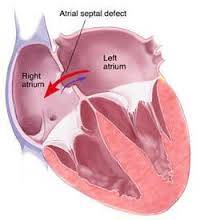
Atrial septal defect (ASD) is a congenital heart defect in which blood flows between the atria (upper chambers) of the heart. Normally, the atria are separated by a dividing wall, the interatrial septum. If this septum is defective or absent, then oxygen-rich blood can flow directly from the left side of the heart to mix with the oxygen-poor blood in the right side of the heart, or vice versa.  This can lead to lower-than-normal oxygen levels in the arterial blood that supplies the brain, organs, and tissues. However, an ASD may not produce noticeable signs or symptoms, especially if the defect is small. A "shunt" is the presence of a net flow of blood through the defect, either from left to right or right to left. The amount of shunting present, if any, determines the hemodynamic significance of the ASD. A "right-to-left-shunt" typically poses the more dangerous scenario.

Signs and Symptoms

Atrial septal defect signs and symptoms may include:
  • Shortness of breath, especially when exercising
  • Fatigue
  • Swelling of legs, feet or abdomen
  • Heart palpitations or skipped beats
  • Frequent lung infections
  • Stroke
  • Heart murmur, a whooshing sound that can be heard through a stethoscope

Causes

An atrial septal defect (ASD) allows freshly oxygenated blood to flow from the left upper chamber of the heart (left atrium) into the right upper chamber of the heart (right atrium). There, it mixes with deoxygenated blood and is pumped to the lungs, even though it's already refreshed with oxygen.
If the atrial septal defect is large, this extra blood volume can overfill the lungs and overwork the heart. If not treated, the right side of the heart eventually enlarges and weakens. If this process continues, the blood pressure in your lungs increases as well, leading to pulmonary hypertension.
Atrial septal defects can be several types, including:
  • Secundum. This is the most common type of ASD, and occurs in the middle of the wall between the atria (atrial septum).
  • Primum. This defect occurs in the lower part of the atrial septum, and may occur with other congenital heart problems.
  • Sinus venosus. This rare defect occurs in the upper part of the atrial septum.
  • Coronary sinus. In this rare defect, part of the wall between the coronary sinus — which is part of the vein system of the heart — and the left atrium is missing.

Risk Factors

It's not known why atrial septal defects occur, but congenital heart defects appear to run in families and sometimes occur with other genetic problems, such as Down syndrome. If you have a heart defect, or you have a child with a heart defect, a genetic counselor can estimate the odds that any future children will have one.
Some conditions that you have or that occur during pregnancy may increase your risk of having a baby with a heart defect, including:
  • Rubella infection. Becoming infected with rubella (German measles) during the first few months of your pregnancy can increase the risk of fetal heart defects.
  • Drug, tobacco or alcohol use, or exposure to certain substances. Use of certain medications, tobacco, alcohol or drugs, such as cocaine, during pregnancy can harm the developing fetus.
  • Diabetes or lupus. If you have diabetes or lupus, you may be more likely to have a baby with a heart defect.
  • Obesity. Being extremely overweight (obese) may play a role in increasing the risk of having a baby with a birth defect.
  • Phenylketonuria (PKU). If you have PKU and aren't following your PKU meal plan, you may be more likely to have a baby with a heart defect.

Complications

A small atrial septal defect may never cause any problems. Small atrial septal defects often close during infancy. Larger defects can cause mild to life-threatening problems, including:
  • Right-sided heart failure
  • Heart rhythm abnormalities (arrhythmias)
  • Increased risk of a stroke
 Less common serious complications may include:
  • Pulmonary hypertension. If a large atrial septal defect goes untreated, increased blood flow to your lungs increases the blood pressure in the lung arteries (pulmonary hypertension).
  • Eisenmenger syndrome. In rare cases, pulmonary hypertension can cause permanent lung damage. This complication, called Eisenmenger syndrome, usually develops over many years and occurs only in a small percentage of people with large atrial septal defects.

Treatments

Many atrial septal defects close on their own during childhood. For those that don't close, some small atrial septal defects don't cause any problems and may not require any treatment. But many persistent atrial septal defects eventually require surgery to be corrected.  Once someone is found to have an atrial septal defect, a determination of whether it should be corrected has to be made.  Methods of closure of an ASD include surgical closure and percutaneous closure. Surgical mortality due to closure of an ASD is lowest when the procedure is performed prior to the development of significant pulmonary hypertension. The lowest mortality rates are achieved in individuals with a pulmonary artery systolic pressure of less than 40 mm Hg.  

NCLEX National Exam Courses

Overview

  • Elite Reviews Offers A Variety Of Online Courses That Will More Than Adequately Help Prepare The Graduate Nurse To Pass The National Exam.
  • Each Course Includes Sample Questions & The Most Current NCLEX Exam Updates.
NCLEX Free Trial
  • FREE Sample Lecture & Practice Questions
  • Available For 24 Hrs After Registration
  • Click The Free Trial Link To Get Started - NCLEX Free Trial


How It Works

How The Course Works

  • First - Purchase The Course By Clicking On The Blue Add To Cart Button - You Will Then Be Prompted To Create A User Account.
  • Second - After Creating An Account, All 3 Options (90, 120, 150 Days) Will Be Listed. Select The Option You Desire And Delete The Other Two.
  • Third - You Will Be Prompted To Pay For This Review Using PayPal - After Payment You Will Be Redirected Back To Your Account.
  • Last - Click The Start Button Located Within Your Account To Begin The Course

NCLEX Predictor Exam

NCLEX Predictor Exam

  • 175 Prep Questions
  • Q & A With Rationales
  • Alt. Format Questions
  • 90 Days Availability
  • Cost $75.00


NCLEX Question Bank

NCLEX Question Bank

  • 1250+ Prep Questions
  • Q & A With Rationales
  • Alt. Format Questions
  • 90 Days Availability
  • Cost $200.00


NCLEX Practice Questions

NCLEX Practice Questions Bundle

  • 1350+ Prep Questions
  • Q & A With Rationales
  • Alt. Format Questions
  • 90 Days Availability
  • Cost $225.00


NCLEX Review

NCLEX Review Course

  • Option 1
  • Lectures & 1250+ Questions
  • Q & A With Rationales
  • Alt. Format Questions
  • 90 Days Availability
  • Cost $275.00


NCLEX Online Review

NCLEX Online Review

  • Option 2
  • Lectures & 2000+ Questions
  • Q & A With Rationales
  • Alt. Format Questions
  • 90 Days Availability
  • Cost $325.00


NCLEX Online Review

NCLEX Review Course Bundle

  • Option 3
  • Lectures & 3000+ Questions
  • Q & A With Rationales
  • Alt. Format Questions
  • 90 Days Availability
  • Cost $375.00


Share On Social Media

Share On Social Media
 Connect With Us - Facebook - Instagram - Twitter 
Read more...

NCLEX Muscular Dystrophy

 NCLEX Muscular Dystrophy Review

NCLEX Muscular Dystrophy

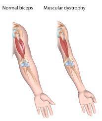
Muscular dystrophy (MD) is a group of muscle diseases that weaken the musculoskeletal system and hamper locomotion (walking or moving ).  Muscular dystrophies are characterized by progressive skeletal muscle weakness, defects in muscle proteins, and the death of muscle cells and tissue.

Signs and Symptoms

The main sign of muscular dystrophy is progressive muscle weakness. Specific signs and symptoms begin at different ages and in different muscle groups, depending on the type of muscular dystrophy.

Duchenne muscular dystrophy

About half of people with muscular dystrophy have this variety. Although girls can be carriers and mildly affected, the disease typically affects boys.
About one-third of boys with Duchenne muscular dystrophy don't have a family history of the disease, possibly because the gene involved may be subject to sudden abnormal change (spontaneous mutation).
Signs and symptoms typically appear between the ages of 2 and 3, and may include:
  • Frequent falls
  • Difficulty getting up from a lying or sitting position
  • Trouble running and jumping
  • Waddling gait
  • Walking on the toes
  • Large calf muscles
  • Muscle pain and stiffness
  • Learning disabilities

Causes

These conditions are generally inherited, and the different muscular dystrophies follow various inheritance patterns. Muscular dystrophy can be inherited by individuals as an X-linked disorder, a recessive or dominant disorder. Furthermore, it can be a spontaneous mutation which means errors in the replication of DNA and spontaneous lesions. Spontaneous lesions are due to natural damage to DNA, where the most common are depurination and deamination

Risk Factors

Muscular dystrophy occurs in both sexes and in all ages and races. However, the most common variety, Duchenne, usually occurs in young boys. People with a family history of muscular dystrophy are at higher risk of developing the disease or passing it on to their children.

Complications

The complications of progressive muscle weakness include:
  • Inability to walk. Some people with muscular dystrophy eventually need to use a wheelchair.
  • Shortening of muscles or tendons around joints (contractures). Contractures can further limit mobility.
  • Breathing problems. Progressive weakness can affect the muscles associated with breathing. People with muscular dystrophy may eventually need to use a breathing assistance device (ventilator), initially at night but possibly also in the day.
  • Curved spine (scoliosis). Weakened muscles may be unable to hold the spine straight.
  • Heart problems. Muscular dystrophy can reduce the efficiency of the heart muscle.
  • Swallowing problems. If the muscles involved with swallowing are affected, nutritional problems and aspiration pneumonia may develop. Feeding tubes may be an option.

Treatments

Currently, there is no cure for muscular dystrophy. Medications and various therapies help slow the progression of the disease and keep the patient mobile for the longest possible time.

Drugs

The two most commonly prescribed drugs for muscular dystrophy are:
  • Corticosteroids: although this type of medication can help increase muscle strength and slow progression, their long-term use can weaken bone and increase weight gain
  • Heart medications: if the muscular dystrophy impacts the heart, beta blockers and angiotensin-converting enzyme (ACE) inhibitors may be useful.

Physical therapy

  • General exercises: a range of motion and stretching exercises can help combat the inevitable inward movement of the limbs as muscles and tendons shorten. Limbs tend to become fixed in position and these types of activities can help keep them mobile for longer. Standard low-impact aerobic exercises such as walking and swimming can also help slow the disease's progression
  • Breathing assistance: as the muscles charged with breathing become weaker, it may be necessary to use devices to help improve oxygen delivery through the night. In the most severe cases, a patient may need to use a ventilator to breathe on their behalf
  • Mobility aids: canes, wheelchairs and walkers
  • Braces: these keep muscles and tendons stretched and help slow their shortening. They also give added support to the user when moving.
 

NCLEX National Exam Courses

Overview

  • Elite Reviews Offers A Variety Of Online Courses That Will More Than Adequately Help Prepare The Graduate Nurse To Pass The National Exam.
  • Each Course Includes Sample Questions & The Most Current NCLEX Exam Updates.
NCLEX Free Trial
  • FREE Sample Lecture & Practice Questions
  • Available For 24 Hrs After Registration
  • Click The Free Trial Link To Get Started - NCLEX Free Trial


How It Works

How The Course Works

  • First - Purchase The Course By Clicking On The Blue Add To Cart Button - You Will Then Be Prompted To Create A User Account.
  • Second - After Creating An Account, All 3 Options (90, 120, 150 Days) Will Be Listed. Select The Option You Desire And Delete The Other Two.
  • Third - You Will Be Prompted To Pay For This Review Using PayPal - After Payment You Will Be Redirected Back To Your Account.
  • Last - Click The Start Button Located Within Your Account To Begin The Course

NCLEX Predictor Exam

NCLEX Predictor Exam

  • 175 Prep Questions
  • Q & A With Rationales
  • Alt. Format Questions
  • 90 Days Availability
  • Cost $75.00


NCLEX Question Bank

NCLEX Question Bank

  • 1250+ Prep Questions
  • Q & A With Rationales
  • Alt. Format Questions
  • 90 Days Availability
  • Cost $200.00


NCLEX Practice Questions

NCLEX Practice Questions Bundle

  • 1350+ Prep Questions
  • Q & A With Rationales
  • Alt. Format Questions
  • 90 Days Availability
  • Cost $225.00


NCLEX Review

NCLEX Review Course

  • Option 1
  • Lectures & 1250+ Questions
  • Q & A With Rationales
  • Alt. Format Questions
  • 90 Days Availability
  • Cost $275.00


NCLEX Online Review

NCLEX Online Review

  • Option 2
  • Lectures & 2000+ Questions
  • Q & A With Rationales
  • Alt. Format Questions
  • 90 Days Availability
  • Cost $325.00


NCLEX Online Review

NCLEX Review Course Bundle

  • Option 3
  • Lectures & 3000+ Questions
  • Q & A With Rationales
  • Alt. Format Questions
  • 90 Days Availability
  • Cost $375.00


Share On Social Media

Share On Social Media
 Connect With Us - Facebook - Instagram - Twitter 
Read more...

NCLEX Myasthenia Gravis

 NCLEX Myasthenia Gravis Review

NCLEX Myasthenia Gravis

Myasthenia gravis (MG) is a neuromuscular disease that leads to fluctuating muscle weakness and fatigue. In the most common cases, muscle weakness is caused by circulating antibodies that block nicotinic acetylcholine receptors at the post-synaptic neuromuscular junction. By blocking the ability of the neurotransmitter acetylcholine to bind to these receptors in the muscle, these antibodies keep motor neurons from signaling the muscle to contract. Alternatively, in a much rarer form, muscle weakness is caused by a genetic defect in some portion of the neuromuscular junction that is inherited at birth as opposed to developing through passive transmission from the mother's immune system at birth or through autoimmunity later in life. Myasthenia gravis is believed to be caused by variations in certain genes. The disorder occurs when the immune system malfunctions and attacks the body's tissues. The antibody in myasthenia gravis attacks normal human protein, targeting a protein called an acetylcholine receptor, or a related protein called a muscle-specific kinase. Human leukocyte antigens have been associated with MG susceptibility.  Many of these genes are present among other autoimmune diseases. Relatives of MG patients have a higher percentage of other immune disorders. The thymus gland cells form part of the body's immune system. In those with myasthenia gravis, the thymus gland is large and abnormal. It sometimes contains clusters of immune cells which indicate lymphoid hyperplasia, and it is believed the thymus gland may give wrong instructions to immune cells

Signs and Symptoms

Muscle weakness caused by myasthenia gravis worsens as the affected muscle is used repeatedly. Because symptoms usually improve with rest, your muscle weakness may come and go. However, myasthenia gravis symptoms tend to progress over time, usually reaching their worst within a few years after the onset of the disease.
Although myasthenia gravis can affect any of the muscles that you control voluntarily, certain muscle groups are more commonly affected than others.

Eye muscles

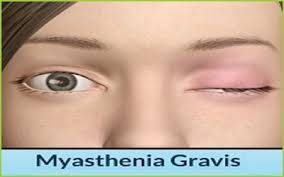
In more than half the people who develop myasthenia gravis, their first signs and symptoms involve eye problems, such as:
  • Drooping of one or both eyelids (ptosis).
  • Double vision (diplopia), which may be horizontal or vertical, and improves or resolves when one eye is closed.

Face and throat muscles

In about 15 percent of people with myasthenia gravis, the first symptoms involve face and throat muscles, which can cause:
  • Altered speaking. Your speech may sound very soft or nasal, depending upon which muscles have been affected.
  • Difficulty swallowing. You may choke very easily, which makes it difficult to eat, drink or take pills. In some cases, liquids you're trying to swallow may come out your nose.
  • Problems chewing. The muscles used for chewing may wear out halfway through a meal, particularly if you've been eating something hard to chew, such as steak.
  • Limited facial expressions. Your family members may comment that you've "lost your smile" if the muscles that control your facial expressions have been affected.

Neck and limb muscles

Myasthenia gravis can cause weakness in your neck, arms and legs, but this usually happens along with muscle weakness in other parts of your body, such as your eyes, face or throat. The disorder usually affects arms more often than legs. However, if it affects your legs, you may waddle when you walk. If your neck is weak, it may be hard to hold up your head.

Causes

This neuromuscular disease is caused by transmission defects in nerve impulses to muscles. The neuromuscular junction is apparently affected: acetylcholine, which produces muscle contraction under normal conditions no longer produces the contractions necessary to muscle movement.[

Treatments

Treatment is by medication and/or surgery. Medication consists mainly of acetylcholinesterase inhibitors to directly improve muscle function and immunosuppressant drugs to reduce the autoimmune process. Thymectomy is a surgical method to treat MG.

Medications

  • Cholinesterase inhibitors
  • Corticosteroids
  • Immunosuppressants

Intravenous therapy

  • Plasmapheresis
  • Intravenous immunoglobulin (IVIG).
  • Monoclonal antibody
 

NCLEX National Exam Courses

Overview

  • Elite Reviews Offers A Variety Of Online Courses That Will More Than Adequately Help Prepare The Graduate Nurse To Pass The National Exam.
  • Each Course Includes Sample Questions & The Most Current NCLEX Exam Updates.
NCLEX Free Trial
  • FREE Sample Lecture & Practice Questions
  • Available For 24 Hrs After Registration
  • Click The Free Trial Link To Get Started - NCLEX Free Trial


How It Works

How The Course Works

  • First - Purchase The Course By Clicking On The Blue Add To Cart Button - You Will Then Be Prompted To Create A User Account.
  • Second - After Creating An Account, All 3 Options (90, 120, 150 Days) Will Be Listed. Select The Option You Desire And Delete The Other Two.
  • Third - You Will Be Prompted To Pay For This Review Using PayPal - After Payment You Will Be Redirected Back To Your Account.
  • Last - Click The Start Button Located Within Your Account To Begin The Course

NCLEX Predictor Exam

NCLEX Predictor Exam

  • 175 Prep Questions
  • Q & A With Rationales
  • Alt. Format Questions
  • 90 Days Availability
  • Cost $75.00


NCLEX Question Bank

NCLEX Question Bank

  • 1250+ Prep Questions
  • Q & A With Rationales
  • Alt. Format Questions
  • 90 Days Availability
  • Cost $200.00


NCLEX Practice Questions

NCLEX Practice Questions Bundle

  • 1350+ Prep Questions
  • Q & A With Rationales
  • Alt. Format Questions
  • 90 Days Availability
  • Cost $225.00


NCLEX Review

NCLEX Review Course

  • Option 1
  • Lectures & 1250+ Questions
  • Q & A With Rationales
  • Alt. Format Questions
  • 90 Days Availability
  • Cost $275.00


NCLEX Online Review

NCLEX Online Review

  • Option 2
  • Lectures & 2000+ Questions
  • Q & A With Rationales
  • Alt. Format Questions
  • 90 Days Availability
  • Cost $325.00


NCLEX Online Review

NCLEX Review Course Bundle

  • Option 3
  • Lectures & 3000+ Questions
  • Q & A With Rationales
  • Alt. Format Questions
  • 90 Days Availability
  • Cost $375.00


Share On Social Media

Share On Social Media
 Connect With Us - Facebook - Instagram - Twitter 
Read more...